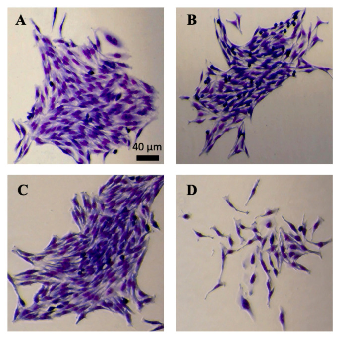
FIGURE 1.
Micrographs of HeLa cell colonies grown under different conditions. Clonogenic assays were performed using six-well plates. Cells were grown in DMEM supplemented with 10% FBS (A), 5% FBS (B), 10% FBS and DMSO (C), or 10% FBS and 60 μM fisetin (D). All images were acquired after HeLa cell colonies were fixed, stained with eosin dye and then methylene dye, and rinsed with water. Scale bar, 40 μm.

