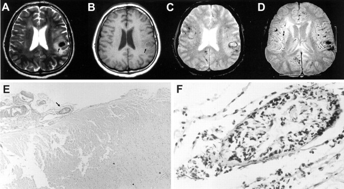
Fig 1.
Images obtained in a 42-year-old man who presented with a history of three stereotypic tingling spells in the right hand.
A, Turbo spin-echo T2-weighted image, obtained through the level of lateral ventricles, shows an acute hematoma with surrounding edema (arrow) in the suprasylvian subcortical region on the left. Additional periventricular hyperintense lesions can be seen, possibly representing ischemic changes of indeterminate age.
B, Corresponding T1-weighted image also shows an acute hematoma with surrounding edema (arrow) in the suprasylvian subcortical region on the left.
C, Gradient-echo T2-weighted image, obtained through the same level as that of the images shown in A and B, shows additional millimetric hypointense foci, consistent with chronic hemorrhages.
D, Gradient-echo image (520/12.9 [TR/TE]; flip angle, 30 degrees), obtained through the level of the basal ganglia, clearly shows multiple hypointense foci of chronic hemorrhages located in the cortical-subcortical region in both hemispheres, with a widespread distribution, whereas the deep gray matter appears to be spared.
E, Pathologic examination of the biopsy material reveals leptomeningeal fibrosis and scattered lymphocytic infiltration with destruction of the vessel wall (arrow) (hematoxylin and eosin, original magnification ×40). Also note petechial hemorrhages with predilection around vessels (arrowheads).
F, Higher magnification of a leptomeningeal blood vessel shows severe destruction of the wall with a mixed inflammatory cell infiltrate (original magnification, ×400).

