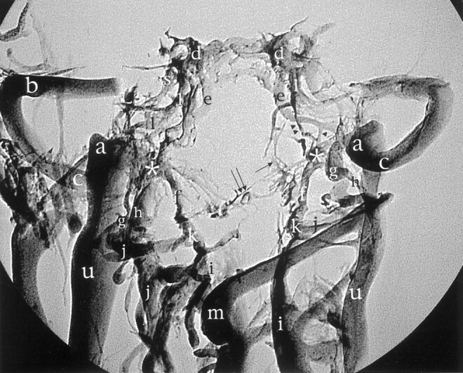
Fig 7.

Standard radiographic picture of the corrosion cast from Figure 2 in the anteroposterior projection shows the ACC (asterisk) and its relation with surrounding veins. The proximal portions of the transverse sinus (b), confluens sinuum, and straight and superior longitudinal sinuses have been removed for better visualization. Arrowhead, branch between the inferior petrosal sinus and the ACC; double arrowhead, inferior petrooccipital vein; arrow, basilar plexus; double arrow, branch to prevertebral venous plexus; d, cavernous sinus; e, inferior petrosal sinus; l, internal carotid artery venous plexus of Rektorzik; a, superior jugular bulb; g, posterior condylar vein; c, sigmoid sinus; h, lateral condylar vein; k, anastomosis between anterior internal vertebral venous plexus and vertebral artery venous plexus; j, vertebral artery venous plexus; u, internal jugular vein; i, anterior internal vertebral venous plexus; m, deep cervical vein.
