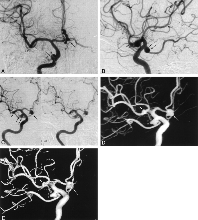
Fig 1.

Images from the case of a 64-year-old female patient with multiple aneurysms.
A, Anteroposterior standard 2D DSA shows right anterior (arrow) and posterior communicating (double arrows) artery aneurysms, but the right middle cerebral artery aneurysm cannot be visualized (arrowhead).
B, Lateral standard 2D DSA shows right anterior (arrow) and posterior communicating (double arrows) artery aneurysms, but the right middle cerebral artery aneurysm cannot be visualized (arrowhead).
C, Rotational DSA image, which can be seen stereoscopically, clearly shows the relationship of the right anterior (arrow) and posterior communicating (double arrows) artery aneurysms to the neighboring vessels and to the aneurysmal necks. The right middle cerebral artery aneurysm (arrowhead) can be seen, but the relationship to the neighboring vessels and the aneurysmal neck are obscured by the superimposition of the surrounding arteries. Note that minimal misregistrations are observed.
D, MIP image clearly shows the relationship of the right anterior (arrow) and posterior communicating (double arrows) artery aneurysms to the neighboring vessels and to the aneurysmal necks. The right middle cerebral artery aneurysm (arrowhead) can be seen, but the relationship to the neighboring vessels and the aneurysmal neck are obscured by the superimposition of the surrounding arteries. Minimal misregistrations do not create artifacts.
E, SSD image clearly shows the relationship of the right anterior (arrow) and posterior communicating (double arrows) artery aneurysms to the neighboring vessels and to the aneurysmal necks. The right middle cerebral artery aneurysm (arrowhead) is seen, and the relationship to the neighboring vessels and aneurysmal neck are easily recognized. Minimal misregistrations do not create artifacts.
