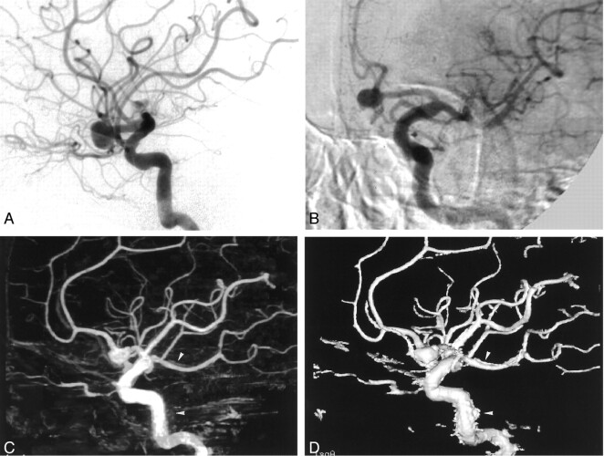
Fig 3.
Images from the case of a 61-year-old male patient with a left anterior communicating artery aneurysm.
A, Lateral standard 2D DSA image. Few image artifacts are noted.
B, Rotational DSA image. Image artifacts are severe.
C, MIP image. Image artifacts create blurring (arrowheads).
D, SSD image. Image artifacts create abnormal irregular structures (arrowheads).

