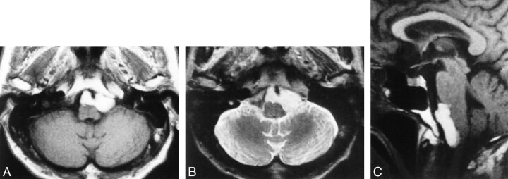
fig 1.
Initial MR imaging.
A, Axial T1-weighted MR image reveals a high-intensity cystic mass, 3.5 cm in diameter, in front of the medulla oblongata.
B, Axial T2-weighted MR image shows that the mass has higher intensity than brain and lower intensity than CSF.
C, Sagittal T1-weighted MR image reveals that the mass compresses the medulla.

