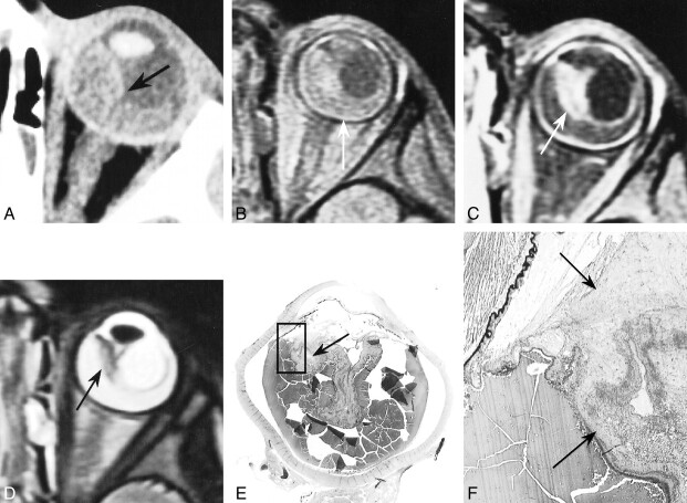
fig 3.
Coats disease in a 2½-year-old boy who presented with strabismus and red eye. Retinal detachment and a whitish mass behind the lens were found on examination of the ocular fundus.
A, Contrast-enhanced CT scan shows large hyperdense detachment (arrow).
B and C, Unenhanced (B) and enhanced (C) T1-weighted MR images show retinal detachment containing material with high signal intensity (arrow, B) and enhancement of the detached retina, forming a mass in the nasal territory (arrow, C).
D, T2-weighted MR image shows the mass has relatively low signal intensity (arrow).
E and F, Low- (F) and high-power (G) histologic sections show Coats disease with retinal detachment, reactive gliosis, and preretinal fibrosis locally constituting a fibrotic block (arrows) containing neovessels (hematein-eosin-saffron stain, original magnifications ×2 and ×25, respectively). (Box in F denotes magnified area in G.)

