
fig 1.
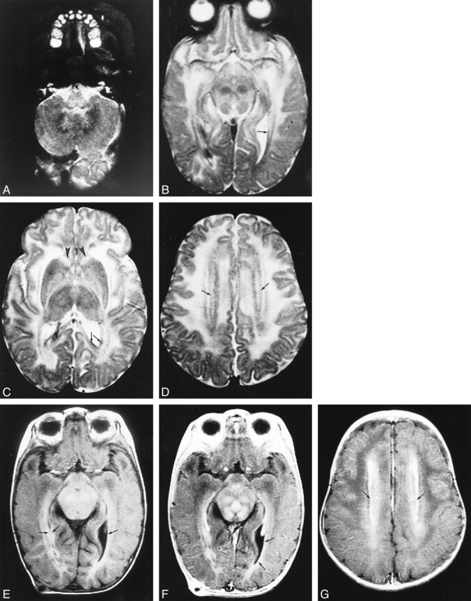
Early MR imaging study at the age of 4 months in a patient with autopsy-proved infantile Alexander disease.
A–D, T2-weighted images show abnormally high signal in the medulla (A), the hilus of the dentate nucleus (arrows, A), the entire midbrain except for the red nuclei (B), the basal ganglia, and the thalamus (C). The frontal white matter has a slightly higher signal intensity than the occipital white matter (C). The head of the caudate nucleus is swollen (arrowheads, C). Around the ventricles, there is a rim of low signal intensity (arrows, B–D).
E–G, T1-weighted images show high signal intensity of the periventricular rim (arrows, E). After contrast administration, the T1-weighted images show enhancement of areas in the midbrain (F), ventricular lining (arrows, F), and periventricular rim (arrows, G).