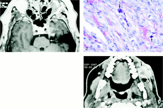
fig 2.
fig 1. Unenhanced axial T1-weighted MR image (728/15/2 [TR/TE/excitations]; matrix, 256 × 512; section thickness, 4 mm) shows an abnormally thickened second division of the trigeminal nerve involving the nerve root entry zone (short arrow) and extending through the prepontine and cavernous (curved arrow) segments.Photomicrograph (hematoxylin and eosin stain) of the original biopsy specimen shows intertwined spindle-shaped cells with pleomorphic hyperchromatic nuclei and frequent mitotic figures (arrows).fig 3. Axial contrast-enhanced CT section obtained through the level of the mandible shows heterogeneously enhancing nodular soft tissue (arrows) within the buccal space. Perineural infiltration along peripheral nerve fibers was confirmed by biopsy.

