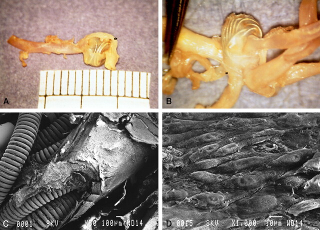
Fig 2.
Gross pathologic findings in case 1.
A, Gross outlook appearance of the thin wall of the aneurysm through which embedded coils are seen.
B, Intraluminal view shows a thin transparent layer of membrane covering the whole orifice of the aneurysm and bridged over the underlying coils.
C, Scanning electron microscopy shows that the coils are covered by thick neointima at the orifice of the aneurysm. Part of the neointima was removed for transmission electron microscopic study (original magnification, ×50; bar = 100 μm).
D, Superficial layer of neointima with a cobblestone appearance (original magnification, ×1000; bar = 10 μm).

