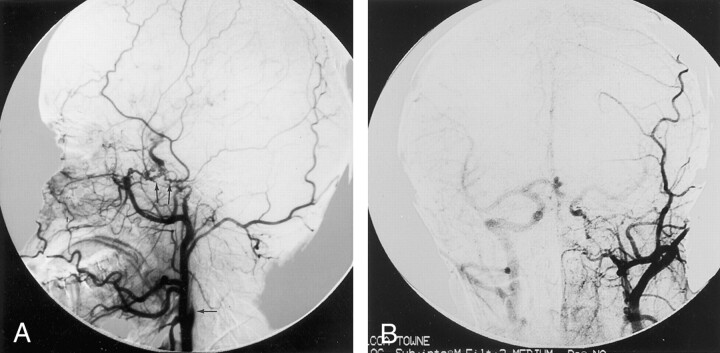
Fig 2.
Left common carotid artery lateral view and Towne injections reveal absence of left ICA (arrow) shortly after its origin and enlarged external carotid artery.
A, Abnormal collateral vessels can be seen between the enlarged left maxillary artery and the supraclinoid segment of the left ICA (arrows).
B, A1 segment of the left anterior cerebral artery is also absent. Right ICA was opacified via backflow from the left common carotid injection.

