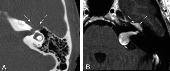
Fig 2.
Axial images demonstrating focal enlargement of the labyrinthine segment of the facial nerve on the axial bone algorithm CT (left), and a homogeneously enhancing mass filling the internal auditory canal with extension into the cerebellopontine angle and labyrinthine segment on the axial T1-weighted postcontrast-enhanced MR image from a similar level (right). These images are from the same case as in Fig 1, and the patient was misdiagnosed preoperatively because of failure to note the enhancement and enlargement along the labyrinthine segment into the geniculate fossa (arrows). From Wiggins and Harnsberger (2001). Used with permission.

