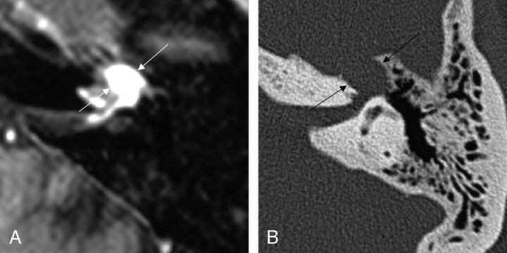
Fig 4.
Axial images from the same case of a facial nerve schwannoma, demonstrating homogeneous enhancement of the labyrinthine segment and geniculate fossa on the axial T1-weighted postcontrast-enhanced MR imaging (left), and focal enlargement of the corresponding geniculate fossa and the labyrinthine segment of the facial nerve on the bone algorithm CT (right), at a similar level (between arrows). From Wiggins and Harnsberger (2001). Used with permission.

