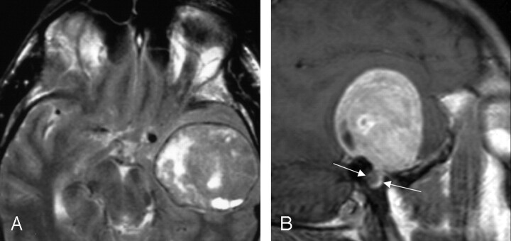
Fig 5.
MR images demonstrating a large left middle cranial fossa mass. The axial T2-weighted image (left), and sagittal T1-weighted postcontrast image (right) show an extra-axial lesion, with a visible CSF/vascular cleft and associated buckling of the gray/white junction. The right image demonstrates a focal bulbous portion of the large mass extending to the geniculate fossa (between arrows), which was the origin of this FNS. From Wiggins and Harnsberger (2001). Used with permission.

