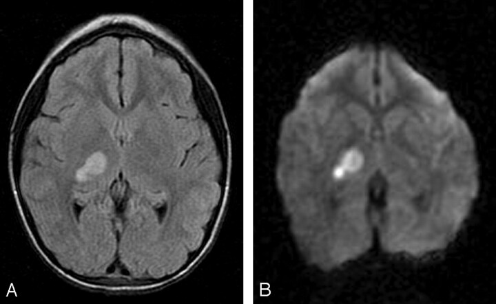
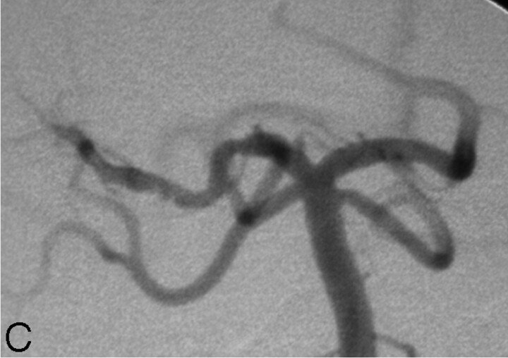
Fig 2.
7-year-old girl presenting with left hemiparesis.
A, Axial FLAIR image demonstrating hyperintensity in the right thalamus and posterior limb of the internal capsule.
B, Axial DWI image showing corresponding restricted diffusion. There was respective low signal intensity on the apparent diffusion coefficient map (not shown).
C, DSA, left vertebral artery injection, transfacial image demonstrating a focal narrowing and irregularity of the proximal right PCA.


