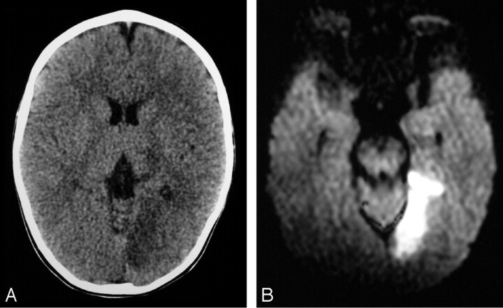
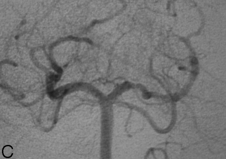
Fig 3.
19-month old boy presenting with right hemiparesis.
A, Axial noncontrast head CT demonstrating hypoattenuation in the medial aspect of the left occipital lobe involving the cortex and subcortical white matter with loss of gray-white matter differentiation.
B, Axial DWI showing corresponding restricted diffusion compatible with infarct. There was respective low signal intensity on the apparent diffusion coefficient map (not shown).
C, DSA, right vertebral artery injection, transfacial image demonstrating an irregular and narrowed left proximal PCA.


