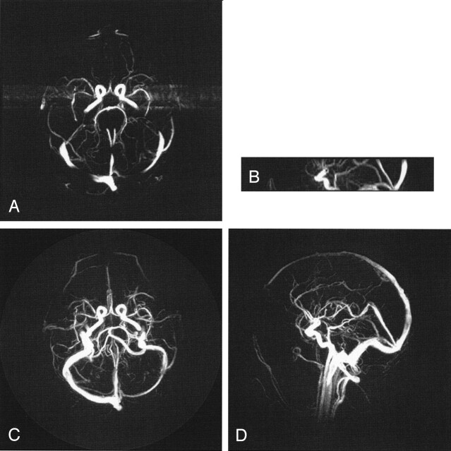
Fig 3.
Comparison of 3DPC (A, B) with a PCVIPR (C, D) acquisition having an acceleration factor of 61. The acceleration is due to a factor of 4.5 in volume coverage, a factor of 7 reduction in voxel volume, and a factor of 2 reduction in imaging time. Visualization of some small vessels is decreased in the PCVIPR examination, which indicates insufficient SNR to support this acceleration factor.

