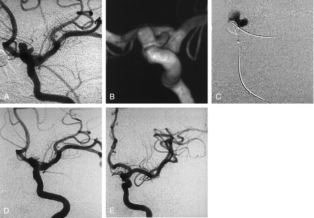
Fig 1.
Incidental aneurysm discovered at MR angiography in a 39-year-old woman with headaches.
A and B, Left ICA conventional (A) and 3D (B) angiograms show a wide-neck bilobed carotid-ophthalmic aneurysm.
C, Road map obtained during balloon inflation and delivery of liquid embolic.
D, Follow-up angiogram obtained immediately after treatment shows complete occlusion of the aneurysm and patency of the ICA.
E, Follow-up angiogram obtained at 24 months shows stable occlusion of the aneurysm.

