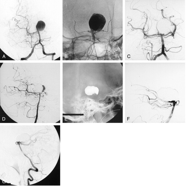
Fig 3.
Basilar artery aneurysm in a 48-year-old woman who presented with symptoms of progressive hemiparesis.
A, Left vertebral angiogram shows a giant basilar tip aneurysm.
B, Nonsubstracted angiogram obtained during inflation of two remodeling balloons and delivery of liquid embolic.
C, Follow-up angiogram obtained immediately after selective endovascular treatment with liquid embolic shows complete occlusion of the aneurysm.
D, Follow-up angiogram obtained at 12 months shows partial recanalization of the aneurysm.
E and F, Follow-up nonsubstracted (E) and substracted (F) angiograms obtained after a second embolization with coils only, show complete occlusion of the aneurysm.
G, Follow-up angiogram obtained at 12 months after second procedure shows persistent complete occlusion of the aneurysm.

