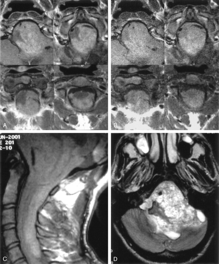
Fig 1.

Contrast-enhanced (A) and nonenhanced (B) T1-weighted axial MR images reveal a posterior fossa mass that extends cranially through 4th ventricle and left cerebellopontine cistern and caudally through foramen magnum. Nonenhanced T1-weighted sagittal MR image (C) shows that a hyperintense melanin-containing mass displaces the medulla oblongata and spinal cord and extends down to the level of the 6th cervical vertebra. T2-weighted axial MR image (D) shows a heterogeneous posterior fossa mass.
