
Fig 1.
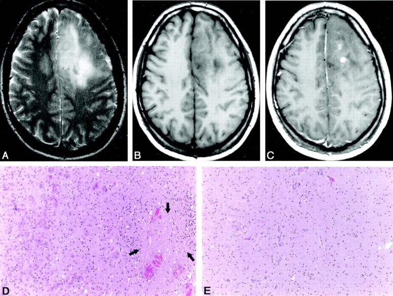
Anaplastic oligodendroglioma obtained in a 28-year-old man.
A, Axial T2-weighted (3588/99 TR/TE) fast SE image shows a high-signal-intensity mass in the left frontal lobe.
B, Axial T1-weighted (540/14) image shows that the mass has low signal intensity.
C, Axial contrast-enhanced T1-weighted (540/14) image shows nodular-like enhancement within the mass.
D, Photomicrograph of the specimen (hematoxylin-eosin stain; original magnification, x25) shows a nodular area that has the high-grade features consisting of high cellularity and nuclear hyperchromasia; however, this is not a specifically sampled area of the nodular-like enhancement on the T1-weighted image. Note the area of necrosis (arrows).
E, Photomicrograph of the specimen (hematoxylin-eosin stain; original magnification, x25) shows the area that has the relatively low-grade features, such as even cellularity and little nuclear pleomorphism. No necrosis is present.