
Fig 2.
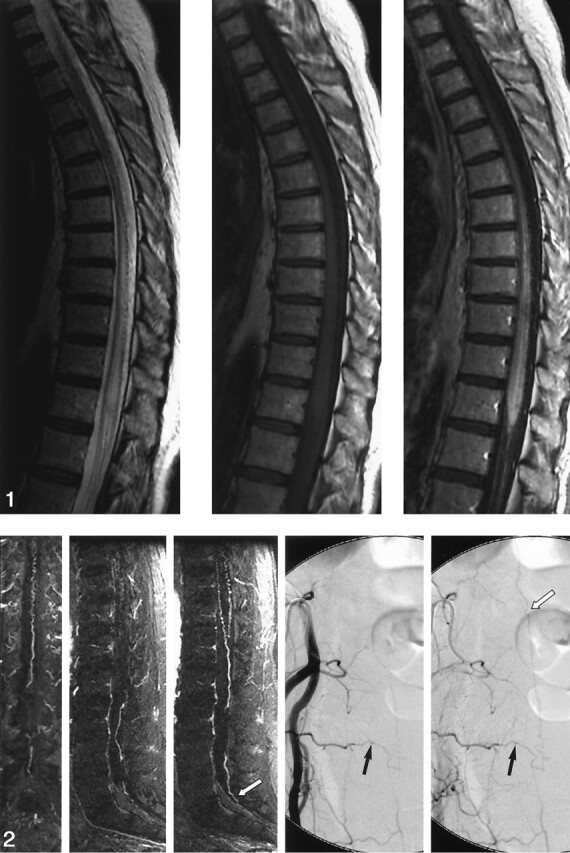
Sagittal MRA performed with a bolus of gadolinium-based contrast agent. Thin, 6-mm coronal (far left) and sagittal (middle left and middle) MIP images obtained at 2-mm increments through the spinal canal show a prominent, tortuous medullary vein extending from the level of the lower body of S1 (white arrow) to the conus and a dilated coronal venous plexus. Catheter, right internal iliac angiograms (middle right and right) show a spinal dural AVF at S4 on the right (black arrows). Draining medullary vein is relatively straight until it begins to meander in the subarachnoid space at the level of the lower body of S1 (white arrow).