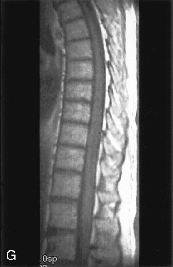
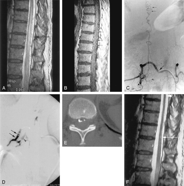
Fig 1.

A, Sagittal T2-weighted MR image cord swelling and central increased signal intensity into the conus, as well as enlarged pial vessels (arrow). B, Sagittal contrast-enhanced T1-weighted MR image shows enhancing enlarged pial vessels (arrows). C, Anterioposterior view of spinal angiogram (injection of intercostal artery) shows filling of an SDAVF with pial drainage (arrowheads); arising from right T9 pedicle (arrow indicates catheter tip). D, Final angiogram control shows embolization cast (arrows) until the initial part of the venous drainage (arrowheads) and no more evidence of the SDAVF or the pial drainage. E, Axial CT control shows clearly embolization cast within the dura mater and into the proximal venous drainage. F, Posttreatment (15 months) sagittal T2-weighted MR image with no more evidence of cord swelling (arrowhead); central increased signal intensity has dramatically decreased, but still remains visible (long arrow). G, Sagittal contrast-enhanced T1-weighted MR image shows no more enhancement of pial vessels previously observed on Figure 1B.

