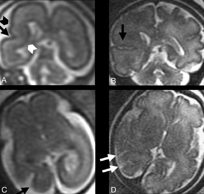
Fig 5.
T2-weighted single-shot, fast spin-echo MR images from the fetal case reported in the “Discussion” section.
A and B, Two coronal sections from studies in gestational weeks 23 and 32, respectively, depicting at right temporal-occipital level the abnormal invagination of a cortical sulcus (black arrow). An additional abnormal smaller sulcus is visible (black arrowhead). The normal migrating cells band is focally thinned (white arrowhead). Note the integrity of ventricular wall, excluding the presence of a schizencephalic cleft.
C and D, Two axial sections from studies in gestational weeks 21 and 32, respectively, showing the early abnormal cortical sulcus (black arrow), followed later on by the development of multiple abnormal sulci (white arrows).

