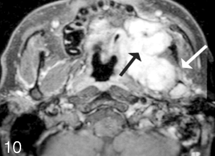
Fig 10.
Sarcoma growing into the left maxilla. Postcontrast fat-suppressed images demonstrate soft-tissue enhancement emanating from the retromolar trigone region (black arrow), with growth into the posterior margin of the left maxilla and abutting the coronoid region of the left mandible. There is likely perineural spread as seen on the axial scan with enhancement into the inferior alveolar canal (white arrow).

