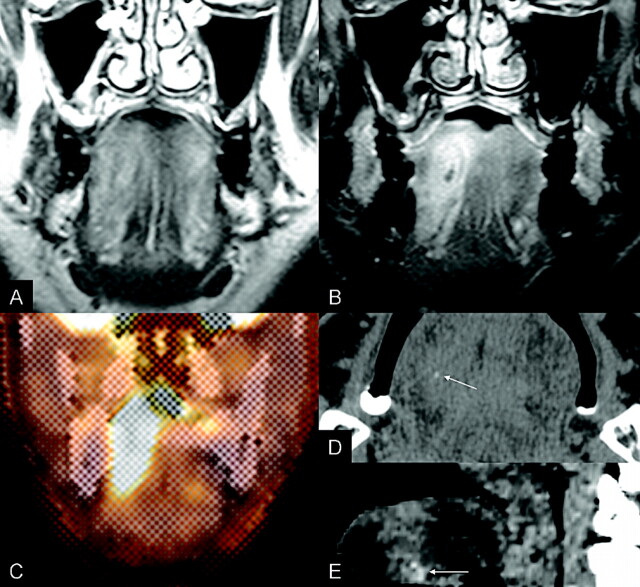
Fig 2.
T2-weighted MR image (A) demonstrates subtle and ill-defined hyperintensity to the right of the middle of the tongue. Gd-enhanced T1-weighted MR image (B) reveals a 4 × 1.6-cm, vertically ovoid, target-like enhancement. The PET-CT image (C) demonstrates a hypermetabolic lesion in the right tongue with a measured peak SUV of 7.4. Noncontrast axial (D) and sagittal reformatted (E) CT images show a small foreign body (arrow) within the slightly hyperattenuated lesion of the anterior tongue.

