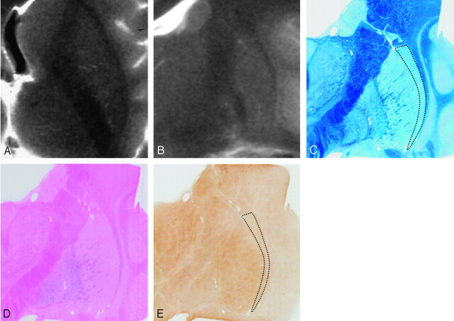
Fig 3.
Comparison of postmortem MR images and histologic findings in a 12-year-old subject. Postmortem axial (A) and coronal (B) T2-weighted images. A section corresponding to B stained with Klüver-Barrera (C), Berlin blue (D), and ferritin immunohistochemistry (E). Postmortem MR images (A and B) show absent HPR. With Klüver-Barrera staining (C), myelinated fibers are small in number in the lateral marginal area of the putamen (dotted lines) in comparison with the remainder. With Berlin blue staining, hemosiderin is scattered in the inner portion of the putamen but is scarce in the outer portion (D). With ferritin immunohistochemistry, ferritin deposits are mild in the inner portion but are scarce in the lateral marginal area of the putamen (dotted lines, E).

