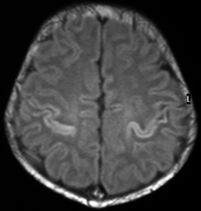
Fig 6.
Term-born infant of a mother with solutio placentae. T1-weighted image (TR/TE, 550/14; signals acquired, 2; matrix, 205 × 256; section thickness, 5 mm; section gap, 0.5 mm; FOV, 16 cm) of the infant from the HIE group at 10 days of age. The image shows higher SI in the PC than in the CR. T1-weighted imaging also showed (not shown here) equal SI in the PP and the PLIC. The flow chart predicted the infant to come from the HIE group.

