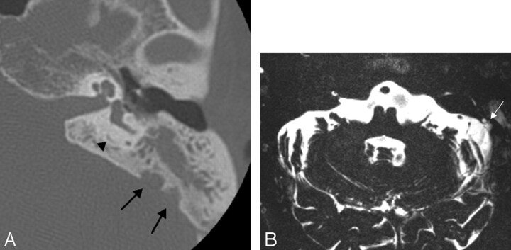
Fig 3.
A, The axial HRCT image of the opacified temporal bone in case 3 shows the presence of an arachnoid granulation (arrows), easily differentiated from an endolymphatic sac tumor by identifying the normal and unenlarged endolymphatic duct (arrowhead).
B, The axial 0.4-mm-thick T2-weighted image confirms the arachnoid granulation (arrow), with CSF signal intensity.

