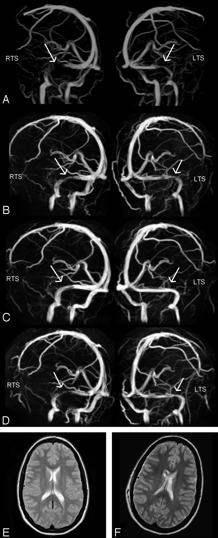
Fig 2.

Case 2: MIP of MRV in oblique anteroposterior left and right views obtained at a lumbar CSF pressures of 50 cm H2O (A), after lumbar puncture (B), after a 2nd lumbar puncture with no residual pressure being monitored (C), and after placement of a ventriculoperitoneal shunt (D). Stenoses in both the left and right transverse sinuses (LTS, RTS), as marked with arrows, are reduced in accordance with a lowered CSF pressure, with complete resolution in C. Probably as a result of a relatively high opening pressure of the valve of the ventriculoperitoneal shunt, transverse sinuses show some residual narrowing (D). T2-weighted axial images before (E) and after (F) insertion of a ventriculoperitoneal shunt into the right ventricle show no difference in ventricle size.
