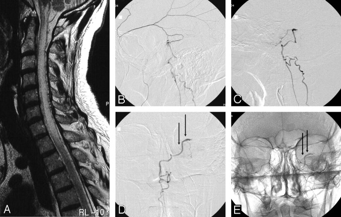
Fig 1.
A 58-year-old man with progressive cervical cord dysfunction.
A, MR image shows a swollen cervical cord with edema and central myelopathy and dilated perimedullary veins.
B, Lateral view of a selective angiogram of the left middle meningeal artery demonstrates a dural fistula, with drainage to the perimedullary veins.
C and D, Lateral (C) and anteroposterior (D) angiograms of the squamous branch of the middle meningeal artery contributing to the fistula show the beginning of the draining vein (arrows). Glue was injected from this position.
E, Anteroposterior radiograph after glue injection shows glue in the draining vein (arrows).

