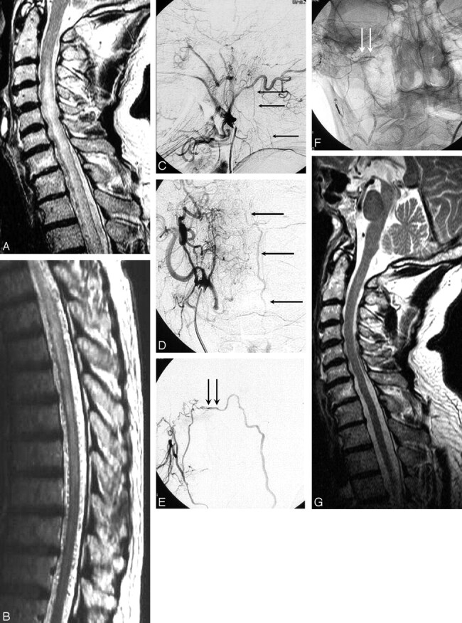
Fig 2.

A 65-year-old man with progressive cord dysfunction.
A and B, MR image shows central myelopathy in the cervicothoracic cord, with engorged perimedullary veins.
C and D, Lateral (C) and anteroposterior (D) right external carotid angiograms demonstrate a fistula with drainage to the perimedullary veins (arrows).
E, Anteroposterior projection of selective injection of a branch of the stylomastoid artery supplying the fistula. The proximal part of draining vein is marked with arrows.
F, Radiograph, same as in E, shows glue in the proximal draining vein (arrows).
G, Normal findings on MR image 1 year later.
