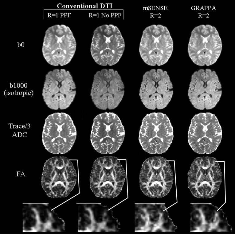
Fig 2.
Image sets (b0, b1000) and Trace/3 ADC and FA maps of a middle section (level of corpus callosum) from one subject with conventional (R = 1), and mSENSE and GRAPPA R = 2 based DTI. Qualitative analysis of the b0 and b1000 images, and Trace/3 ADC maps indicated that mSENSE and GRAPPA R = 2 DTI generated the sharpest images and were more adept at handling spatial warping effects seen in images of the R1-PPF and R1-no PPF methods. Although FA maps derived from R1-PPF were considered to be smoother because of their intrinsically higher SNR in 4 of 5 cases, an in-depth comparison of these maps to those obtained by using mSENSE and GRAPPA R = 2 DTI showed that incorporating parallel imaging allowed better visualization of thinner white matter tracts such as the middle frontal gyrus (magnified, below FA maps).

