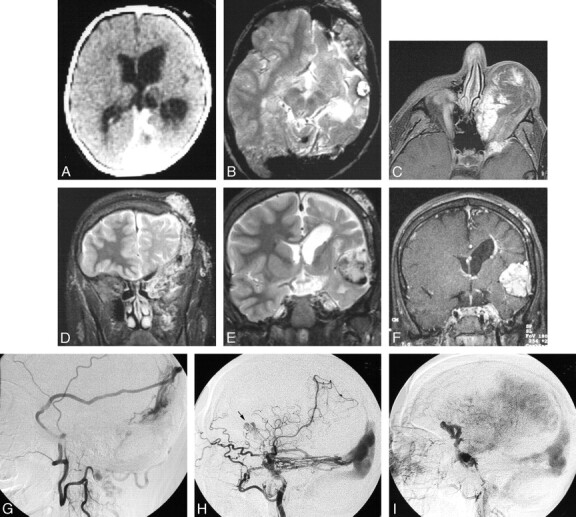
Fig 6.

Serial imaging of a girl with an extensive left orbitofrontal lymphatic malformation associated with a left posterior dural AV fistula, dural sinus enlargement, left cerebral hemiatrophy, sinus pericranii, and progressive formation of cavernoma-like lesions. The patient presented at birth in high output cardiac failure and underwent ligation of numerous extracranial arteries with clinical improvement. She subsequently underwent numerous surgical procedures to treat the orbital LM, which was complicated by recurrent hemorrhage. At age 17, she presented with severe orbital chemosmosis and underwent angiography. It is possible that some of the anterior vascular abnormalities are secondary to the craniotomies performed to debulk the left orbit.
A, Axial CT image after intravenous contrast administration in the first week of life shows the large straight and left transverse sinuses and left hemiatrophy. Note the absence of a focal mass lesion in the left middle cranial fossa.
B, Axial T2-weighted MR imaging of the brain at 10 years of age shows persistent enlargement of the left transverse sinus, and a cavenomalike lesion in the left middle fossa.
C, Axial T1-weighted postgadolinium MR image at 17 years of age, after numerous orbital debulking procedures. There are enhancing channels within the orbital LM and adjacent sphenoid bone. These may represent small arterial lymphatic communications or a pure venous component.
D, Coronal T2-weighted image from the same study as (C) shows LM involvement of the left infratemporal space, left orbit, left sphenoid and frontal bones, and the scalp.
E, Coronal T2-weighted and MR image at the level of the frontal horns demonstrates enlargement of the cavenomalike lesion in the Sylvian fissure.
F, Postgadolinium T1-weighed image shows attenuated enhancement of the extra-axial mass. A periventricular DVA is also present.
G, Left middle meningeal angiogram, lateral projection, (via cervical collateral) shows supply of the dural AVM by the parietal branch of the middle meningeal artery.
H, Left internal carotid angiogram, lateral projection, shows additional supply to the posterior dural AVM by the tentorial branches of the internal carotid artery. Note a second, more anterior lesion (arrow).
I, Venous phase of the left internal carotid angiogram shows opacification of anomalous veins probably constituting part of the vascular mass in the Sylvian fissure.
