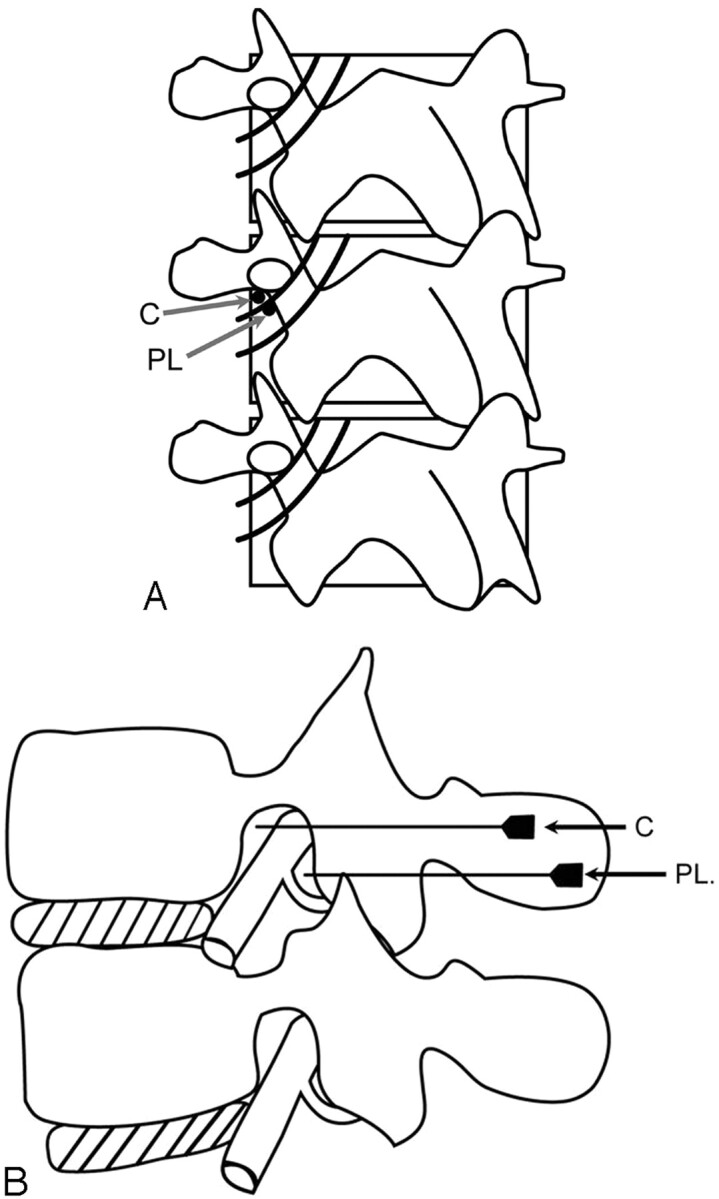
Fig 2.

Schematic description of the conventional and posterolateral TFESI techniques. In oblique view (A), the needle tip is located in the safe triangle using the conventional technique, and the median inferior margin of pedicle with posterolateral approach. The needle appears end-on in this view. Lateral view (B) shows the needle located in the anterior and superior aspect of a nerve root using the conventional technique and at the posterior aspect using the posterolateral technique. C, conventional TFESI; PL, posterolateral TFESI.
