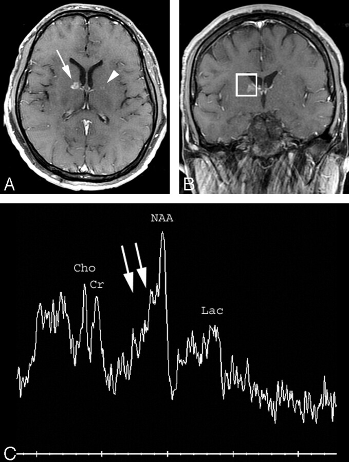
Fig 2.
A, Axial T1-weighted postcontrast MR image (TR/TE = 466/14) shows a small ring-enhancing lesion in the genu of the right internal capsule (arrow) and a barely perceptible lesion in the left globus pallidus (arrowhead). Multiple additional similar small ring-enhancing lesions were identified throughout the brain parenchyma.
B, Voxel localization for proton MR spectroscopy of the right internal capsule lesion.
C, MR spectroscopy of the right internal capsule lesion demonstrates marked elevation of the β,γ-Glx peaks (double arrows) compared with creatine (peak height ratio 1.1 [normal less than 0.5]) compatible with tumefactive multiple sclerosis. There is also mild decrease of N-acetylaspartate and probable mild presence of lactate. NAA indicates N-acetylaspartate; Cho, choline; Cr, creatine; Lac, lactate.

