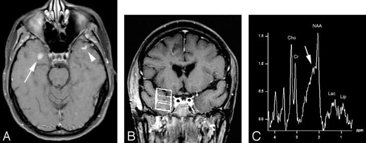
Fig 4.
A, Axial T1-weighted postcontrast MR image (TR/TE = 481/14) shows a solidly enhancing lesion in the right temporal lobe (arrow). There is also a curvilinear focus of enhancement in the left temporal subcortical white matter (arrowhead) suggestive, but not diagnostic, of the diagnosis of demyelinating disease.
B, Voxel localization for proton MR spectroscopy of the right temporal enhancing mass.
C, MR spectroscopy of the right temporal lobe lesion shows mild elevation of choline and mild decrease of N-acetylaspartate metabolites with probable presence of lipid and lactate possibly leading to the incorrect assumption of neoplastic disease. Again, however, there is elevation of the β,γ-Glx peaks (arrow) consistent with the patient’s correct diagnosis of tumefactive multiple sclerosis.

