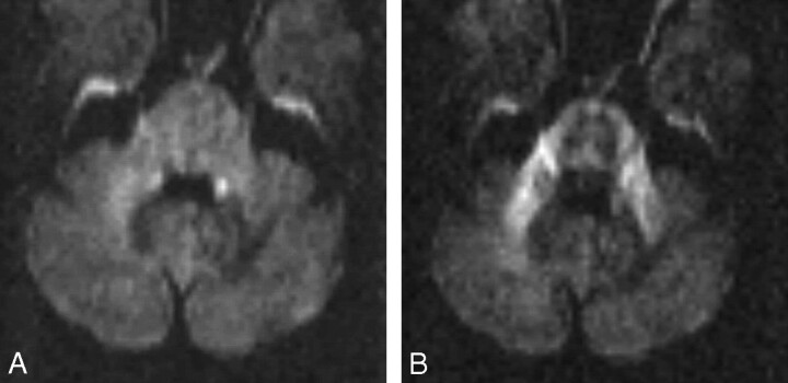
Fig 2.
MR imaging study 4 weeks after commencing HAART.
A, Trace images of DWI3000 no longer show any high-signal-intensity rim at the periphery of the lesion, and the left MCP appears only slightly less bright than the right one.
B, On the anisotropic images of DWI3000, sensitive for water movement in the section (supero-inferior) direction, the left MCP appears now nearly as bright as the right MCP, implying that structural integrity has been regained.

