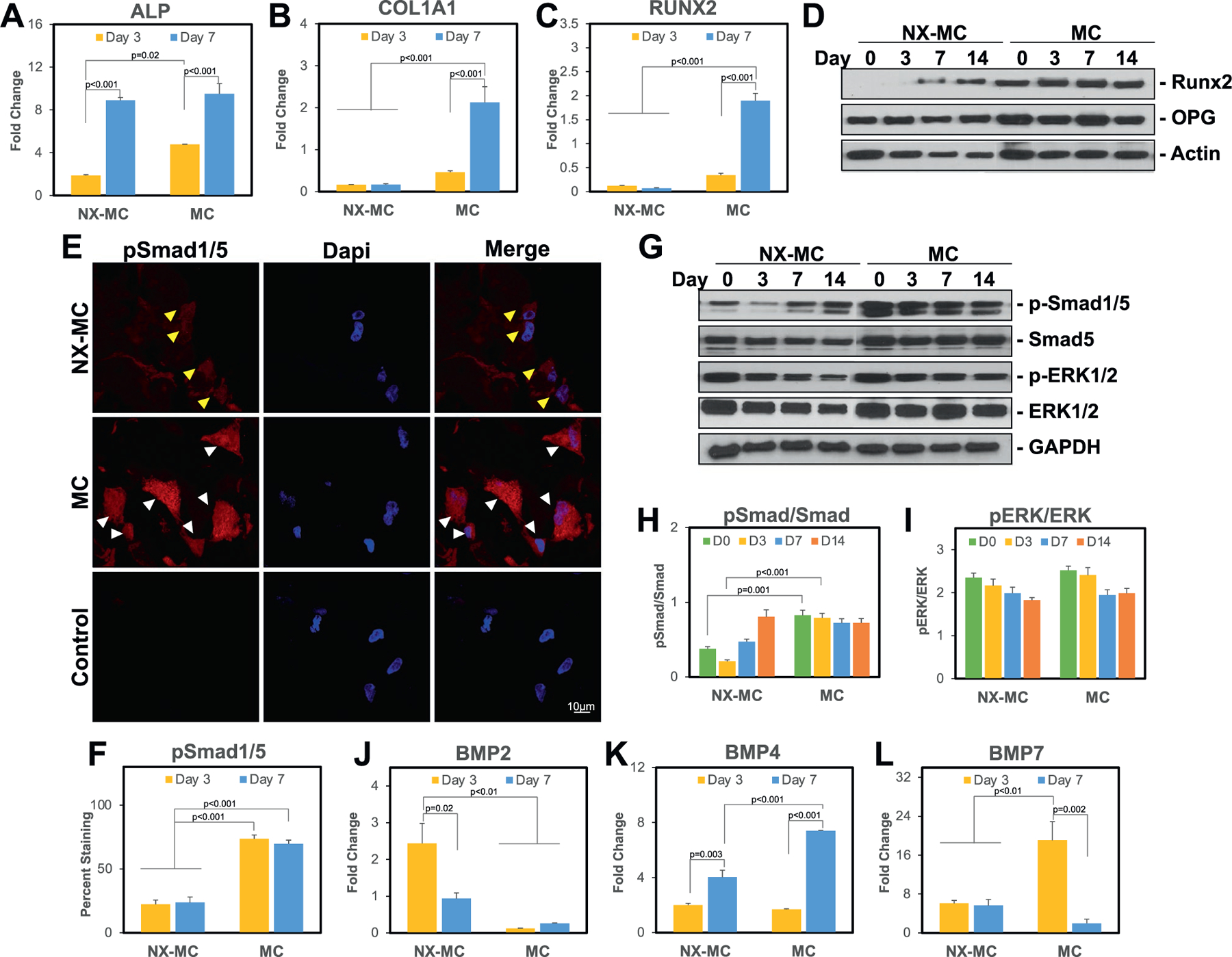
Figure 3.

Stiffness increases expression of osteogenic genes and activation of the canonical BMP receptor and Wnt signaling pathways. QPCR of primary hMSCs cultured on NX-MC or MC for 3 or 7 d in osteogenic differentiation medium for A) ALP, B) COL1A1, C) RUNX2, J) BMP2, K) BMP4, and L) BMP7 (n = 3). E) Representative confocal microscopic images of primary hMSCs cultured on NX-MC (top row) or MC (middle row) for 3 d stained with anti-p-Smad1/5 and Dapi. Negative control (Control) using secondary antibody only and Dapi on cells cultured on MC shown on bottom row. Scale bar indicates 10 µm. Yellow arrows indicate cells with weak p-Smad1/5 staining and sparing of the nuclei. White arrows indicate cells with strong p-Smad1/5 staining including nuclear staining. F) Quantification of p-Smad1/5 staining of primary hMSCs cultured on NX-MC or MC for 3 or 7 d. Western blot of primary hMSCs cultured on NX-MC or MC for 0, 3, 7, or 14 d in osteogenic differentiation medium for D) Runx2, OPG, and β-actin or G) p-Smad1/5, total Smad5, p-ERK1/2, total ERK1/2, and GAPDH. Densitometric quantification of western blot analyses demonstrating relative protein amounts of H) p-Smad1/5 to total Smad5 and I) p-ERK1/2 to total ERK1/2. Bars represent means, errors bars represent SE. Significant posthoc comparisons following ANOVA indicated with p-values.
