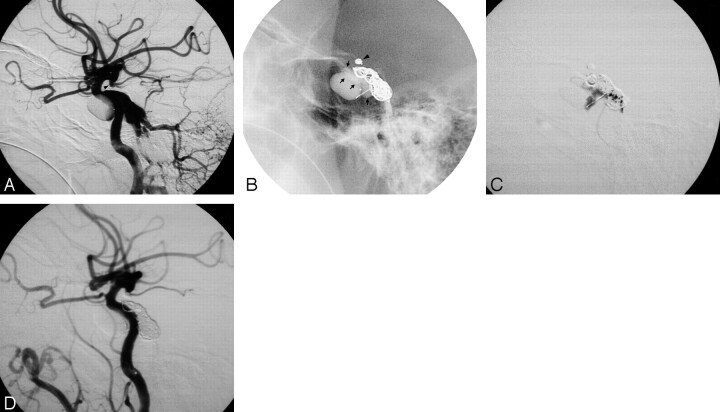
Fig 1.
Images of a 49-year man with a traumatic DCCF and a traumatic aneurysm at the left supraclinoid ICA. A, Left lateral carotid angiogram reveals a residual fistula after a detachable balloon embolization. B and C, Two GDCs were placed into the cavernous sinus. Further coil embolization to achieve angiographic cure failed because of recoil of the microcatheter, and the traumatic aneurysm was occluded by GDC (arrowhead). Under a protective balloon (arrows) at the cavernous portion of the ICA, a total of 0.5 mL of n-BCA mixture was infused into the cavernous sinus. D, Postembolization angiogram reveals total obliteration of the residual fistula with preservation of the ICA flow.

