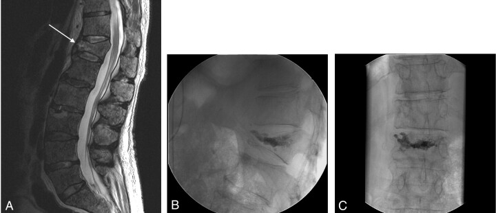
Fig 2.
Prevertebroplasty MR imaging (A) and intraoperative fluoroscopic images (B and C) of the treated level in a patient without an intraosseous cleft. T2-weighted (repetition time [TR]/echo time [TE], 3300/150 fast spin-echo, without fat saturation) MR (A) demonstrates acute compression of T12 in this 61-year-old male patient. Lateral (B) and anteroposterior (C) fluoroscopic images after vertebroplasty at T12 show cement evenly distributed among the trabeculae of the fractured vertebral body.

