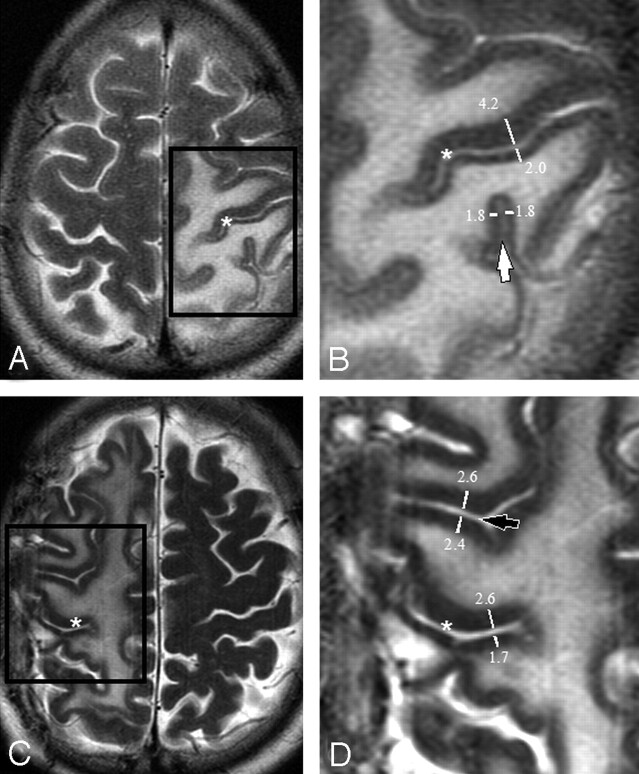
Fig 1.
Axial T2-weighted images showing edema surrounding the central sulcus (asterisks A–D) in 2 patients (A and C). Magnification of the boxed regions (B and D) demonstrate thickness measurements across the central sulcus (asterisks in B and D), across a neighboring parietal sulcus (a ramus of the intraparietal sulcus [white arrow]) and across a neighboring frontal sulcus (precentral sulcus [black arrow]). Only one measurement is shown for clarity. Up to 8 measurements were made along the length of the sulcus on each axial image. Note that measurements were performed perpendicular to the sulcus, ensuring that the ratio will not be affected by section orientation.

