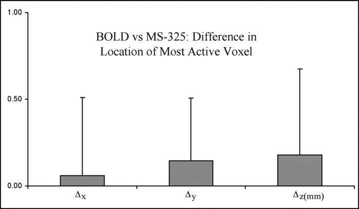
Fig 2.
Average distance (millimeters) between the most active voxel in BOLD fMRI measurements versus the location of the most active voxel in MS-325 fMRI in the same animal (n = 9). The distances (Δx, Δy, and Δz) between the most active voxels were determined for each animal. Data are displayed as mean millimeters ± SD.

