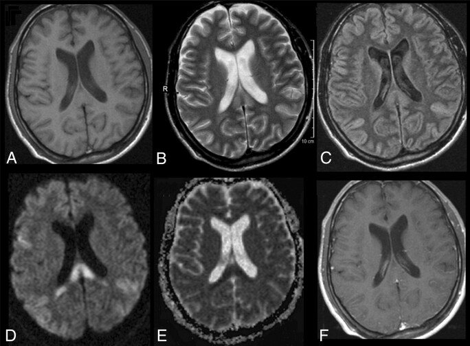
Fig 1.
Initial MR imaging of the patient. T1-weighted image does not reveal any abnormality (A). T2-weighted image depicts high signal intensity in the SCC (B). Turbo spin-echo FLAIR sequence shows additional high signals in both posterior hemispheres (C). All lesions had clearly elevated diffusion coefficients with high signal intensity on DWI (D), whereas ADC maps in the splenium were decreased (E). The T1-weighted image with gadolinium shows no contrast enhancement.

