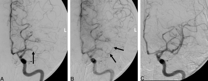
Fig 1.
Prethrombolysis midarterial phase (A) and late arterial phase (B) angiographic images and post-thrombolysis angiographic image (C) from a patient who underwent intra-arterial thrombolysis with complete recanalization (C). This patient presented with a left m1 segment occlusion (A, arrow). Note the delayed opacification of the distal m1 and proximal m2 segments (arrows).

