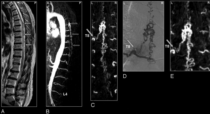
Fig 1.
SDAVF in a 61-year-old patient. Comparison of visualization capabilities of MRA and DSA.
A, Sagittal T2-weighted image showing signal intensity voids raising the suspicion of a vascular spinal cord abnormality (small white arrows).
B, Sagittal MIPs of the MRA examination showing the overview and localization of the dilated vein (small black arrows).
C, In the coronal target MRA MIP the feeding segmental artery of the SDAVF was depicted to derive from the eighth thoracic level (T8) on the left side. Localization of the shunt is at dural level (gray arrow).
DSA (D) provides more spatial resolution and more insight in the dynamic drainage of the dilated vein compared with MRA (E).

