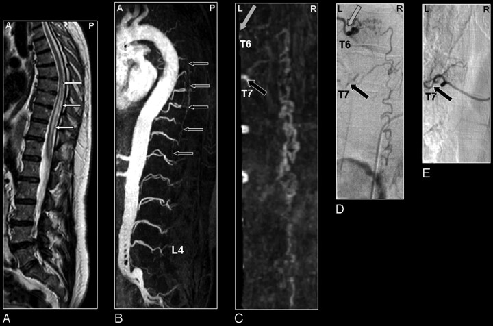
Fig 4.
SDAVF in a 61-year-old male patient visualized by MRA and DSA. Misinterpretation of the segmental level of origin on MRA.
A, Sagittal T2-weighted image showing spinal cord edema (small white arrows).
B, Sagittal MIP of the MRA examination showing the overview and localization of the dilated vein (small black arrows).
C, In the coronal target MRA MIP the feeding segmental artery of the SDAVF was falsely localized by just 1 level at the seventh thoracic level (T7; black arrow).
D, DSA shows that the feeding segmental artery (gray arrow) originates from the sixth thoracic level (T6).
E, Selective injection of segmental artery T7 (black arrow) shows no supply to the SDAVF. Retrospectively, the correct level (gray arrow) could be identified on the target MRA MIP (C).

