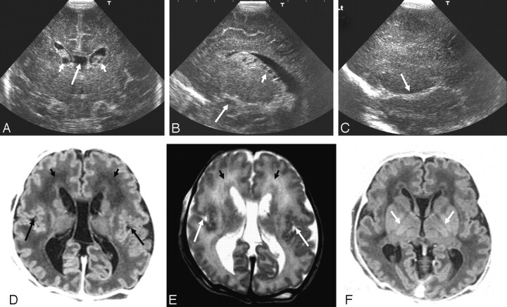
Fig 3.
Ultrasonography (A–C) and MR imaging (D–F) (time interval, 1 day) from an infant with a peroxisomal biogenesis disorder with a Zellweger phenotype.
A, Coronal view showing GLCs (short arrows), a large cavum septum pellucidum (long arrow), and increased echogenicity in the white matter.
B, Parasagittal view showing GLCs, cysts, in the choroid plexus (short arrow) and an abnormal appearance to the insula (long arrow).
C, Extreme parasagittal view showing abnormal development of the Sylvian fissure (arrow) and increased echogenicity in the white matter.
D and E, Axial inversion recovery and T2-weighted MR images showing a large cavum septum pellucidum, abnormal signal intensity in the frontal white matter (short arrows), and polymicrogyria of the Sylvian fissures (long arrows).
F, Axial inversion recovery image showing a lack of myelin in the posterior limb of the internal capsule (arrows).

