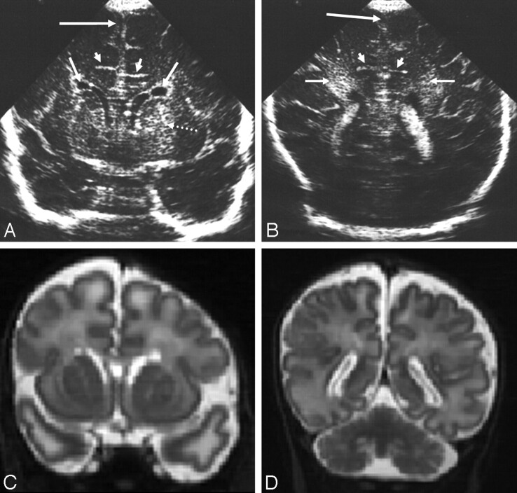
Fig 7.
Ultrasonography (A and B) and MR imaging (C and D) (time interval, 2 days) from an infant with methylmalonic acidaemia.
A, Coronal view showing straight sulci coming off the interhemispheric fissure (short arrows), bilateral GLCs (medium arrows), LSV (dotted arrow), and slightly widened interhemispheric fissure (long arrow).
B, Posterior coronal view showing straight sulci (short arrows), slightly widened interhemispheric fissure and extracerebral space (long arrow), and increased echogenicity in the white matter of the trigone (medium arrows).
C and D, Reconstructed coronal T2-weighted MR images showing features similar to the ultrasonography images except for the LSV (only seen sonographically) and white matter change also seen subcortically on the MR images.

