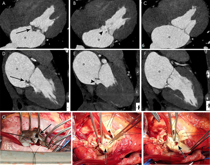
Figure 11:
A–C, Four-chamber and, D–F, two-chamber contrast-enhanced CT images in a 39-year-old woman show focal ill-defined attenuation along the inferior aspect of the anterior mitral valve leaflet (vegetation, arrows) associated with leaflet perforation (arrowheads) and left atrial dilatation (*). G, Intraoperative image in a 32-year-old woman with infective endocarditis shows mitral valve leaflet perforation (arrow). H, I, Intraoperative images in a 49-year-old man with infective endocarditis show aortic valve leaflet perforation (arrows).

